What Is TRIzol and What Is It Used For?
Trizol is a widely used reagent for extracting RNA from cells and tissues. It can also support recovery of DNA and proteins from the same sample, making it useful when researchers need several molecular readouts. The reagent disrupts biological material and helps separate its components during extraction. In their 1987 paper, Piotr Chomczynski and Nicoletta Sacchi described the underlying approach as a “single-step method of RNA isolation by acid guanidinium thiocyanate-phenol-chloroform extraction.” That phrase captures the method’s chemical basis, but not every practical detail.
In this guide, we will explain what TRIzol contains, how its separation process works, and where it is commonly used. We will also cover sample preparation, phase separation, RNA precipitation, and common sources of poor yield or quality. Small choices matter. For example, incomplete homogenization can leave visible tissue fragments and reduce extraction consistency. Clean technique matters, too, because trace contamination may affect downstream analysis. TRIzol is useful, but it is not a shortcut around careful handling. Its performance depends on the sample, the protocol, and the researcher’s judgment. That last point is easy to overlook. Results should be checked with suitable quality controls, and the manufacturer’s instructions and safety information should guide laboratory use. This overview offers a practical starting point, while recognizing that no single protocol fits every sample.
What TRIzol Is: A Single-Step Reagent for RNA, DNA, and Protein Isolation
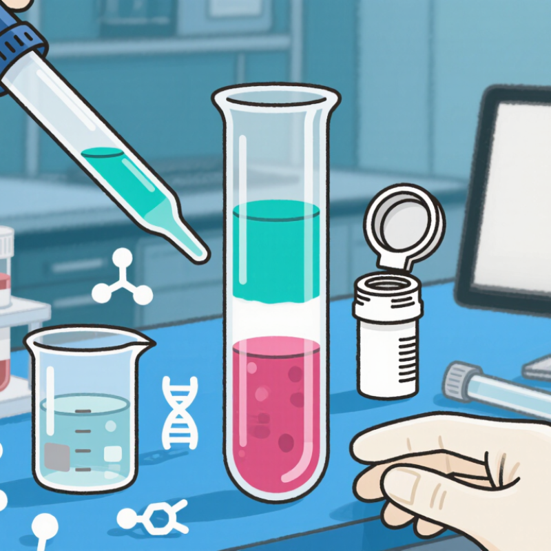
A single-step phenol-guanidinium reagent can isolate RNA, DNA, and proteins from one biological sample. Rather than dividing a small tissue sample among separate extraction workflows, the reagent disrupts cells and helps deactivate enzymes that can degrade RNA. After a phase-separation solvent is added and the sample is centrifuged, distinct layers form. RNA is recovered from the aqueous layer, while DNA and proteins remain in other fractions for separate recovery. Exact results depend on sample type, handling, and protocol details. Small differences matter.
On the bench, tissue is homogenized until visible fragments disappear. After a brief rest and centrifugation, the sample separates into layers that must be collected carefully. A disturbed interface can carry contaminants into the next step. That matters. The method is useful when sample material is limited or several molecular analyses are planned. Yet “single-step” describes the initial disruption, not a hands-off process. Phenol-based reagents and separation solvents require suitable protective equipment, ventilation, and disposal procedures. Follow a validated protocol for the specimen and downstream assay. RNA yield alone does not confirm RNA integrity, and recovered DNA or protein may need additional cleanup. Not quite effortless.
Core Chemistry: Acid Guanidinium–Phenol Extraction (Chomczynski & Sacchi, 1987)
Acid guanidinium–phenol extraction is a single-step method for separating RNA from cells or tissue. Chomczynski and Sacchi described the approach in 1987. Its chemistry combines guanidinium salts, acidic phenol, and a phase-separation step. Guanidinium salts disrupt protein structure and inhibit RNases, enzymes that can quickly degrade RNA. Phenol helps denature proteins and release cellular contents.
After chloroform is added and the mixture is centrifuged, it separates into visible layers. Under acidic conditions, much of the RNA remains in the upper aqueous phase. DNA tends to collect at the interphase, while many proteins partition into the lower organic phase. The boundary can look thin and slightly cloudy. Careful separation matters. A small amount carried across may affect later measurements.
This method is valued because it can recover RNA from complex samples without first isolating individual cells. Yet the separation is not magical: sample composition, mixing, and pH can influence purity and yield. I find that the clean-looking layers can invite overconfidence. The interface is easy to disturb, and downstream checks remain important. Extraction creates a useful starting point, not proof that the RNA is intact or suitable for every experiment.
Input: 1 mL per 50–100 mg Tissue or 5–10 Million Cells (Thermo Fisher)
A phenol–guanidinium extraction reagent is used to break open cells and help preserve RNA during sample processing. The input amount matters: use about 1 mL of reagent for 50–100 mg of tissue, or for 5–10 million cells. These figures are a practical starting range, not a guarantee of yield. Tissue density, cell type, and sample condition can change how easily a sample lyses. A small, firm tissue piece may need more thorough homogenization than the same weight of soft tissue.
Keep the sample fully immersed and distribute the reagent evenly. For tissue, mince it into small pieces before homogenizing; clumps can remain intact and reduce extraction quality. Too much starting material can make the lysate thick and harder to separate cleanly.
That detail is easy to miss. If the sample exceeds the recommended range, divide it between tubes rather than forcing it into one. Record the actual input mass or cell count, since estimates can be imperfect. Handle the reagent with suitable protective equipment and ventilation, and follow your laboratory’s approved waste procedures.
Phase Split: 0.2 mL Chloroform/mL; 12,000 × g, 15 min (Thermo Fisher)
An acidic phenol–guanidinium reagent is used to extract RNA from cells or tissue while disrupting proteins and inactivating many enzymes. Chloroform creates the phase split. For each 1 mL of reagent, add 0.2 mL chloroform, then mix carefully and let the sample stand briefly. Centrifuge at 12,000 × g for 15 minutes. Follow the validated protocol for temperature and handling details.
After centrifugation, the tube should show distinct layers. The upper, clear aqueous phase contains most of the RNA; the interphase and lower organic phase hold much of the DNA and protein. The boundary can look deceptively neat, but it may be faint or uneven. Small volumes matter. Transfer only the aqueous layer, leaving a visible margin above the interphase. A slightly smaller RNA yield is often preferable to carrying contaminants forward.
Tips: Work in a chemical fume hood with suitable gloves and eye protection. Use a pipette with a clean tip, and keep the tube steady while aspirating. Do not rush. If the interface is disturbed, pause and reassess rather than guessing where the layers meet. Centrifugal force is specified as × g, not revolutions per minute; use the rotor’s conversion when needed.
Recovered Biomolecules and Uses: RNA for RT-qPCR, DNA for Genotyping, Protein Analysis
A phenol–guanidinium extraction reagent can separate RNA, DNA, and proteins from the same disrupted cell or tissue sample. After adding a phase-separation solvent and centrifuging, the sample forms distinct layers. RNA is usually recovered from the aqueous layer, while DNA and proteins remain in other fractions. The boundary is not always neat. Careful pipetting helps limit cross-contamination.
Recovered RNA can be reverse-transcribed and measured by RT-qPCR to compare gene expression across samples. Check RNA concentration and integrity, and include suitable controls. Residual reagent can inhibit downstream reactions, even when the RNA looks clean. Small details matter.
DNA recovered from the interphase can support genotyping, provided it is purified and assessed for quality. The protein fraction can be precipitated and prepared for methods such as immunoblotting or other protein analyses. These proteins are denatured, so they are generally unsuitable for studying native activity. It is easy to overtrust a clean-looking pellet; extraction quality still depends on sample handling, fraction recovery, and the assay that follows.


